SonoAnatomy Atlas
Master’s Research Project
Ultrasound (US) imaging is a valuable tool used in many medical disciplines. Currently, there is a lack of standardized US education among Canadian medical schools, and accordingly, limited introductory resources. Challenges such as orienting US planes, translating between 2D images and 3D anatomical relationships, and interpreting structures in grayscale, present a steep learning curve for novice learners. This project aims to enhance undergraduate medical US instruction by creating, evaluating, and sharing an online interactive Sonoanatomy Atlas. The modular atlas covers fundamental US principles, 3D shoulder anatomy, and a shoulder scanning protocol.
-
Michael Corrin and Terry Li
-
Interactive and modular instructional website
-
Autodesk Maya, ZBrush, Adobe Illustrator, Adobe After Effects, Figma
-
Researcher, writer, UX/UI designer, 2D/3D artist, web developer
Open source model of shoulder anatomy, and functional prototype coming soon!
The SonoAnatomy Atlas contains 3 distinct shoulder modules: 1) Anterior shoulder, 2) Posterior shoulder, and 3) Superior shoulder.
Each module integrates user-controlled 2D and 3D components that allow users to interactively explore how US interacts with shoulder anatomy. Instructional animations of the procedural US techniques synchronized with real scans help demonstrate the complex and dynamic anatomy, and colour illustrations of each US scan facilitate development of interpretive skills. We hope to evaluate the resource with students and instructors to assess integration into curriculum, perceived efficacy, website traffic and engagement, and content understanding.
Literature review, media audit, user personas and user journeys, content inventory and script writing!


Wireframing and prototyping in Figma.

3D modelling and rigging.
Throughout the website, the shoulder is examined in 4 different positions; neutral position, external rotation, abduction, modified crass - as well as in transition states.
In order to create orientation images for each step in a scanning protocol, a 3D model of the shoulder anatomy was built and rigged. This model was then positioned and animated as needed.


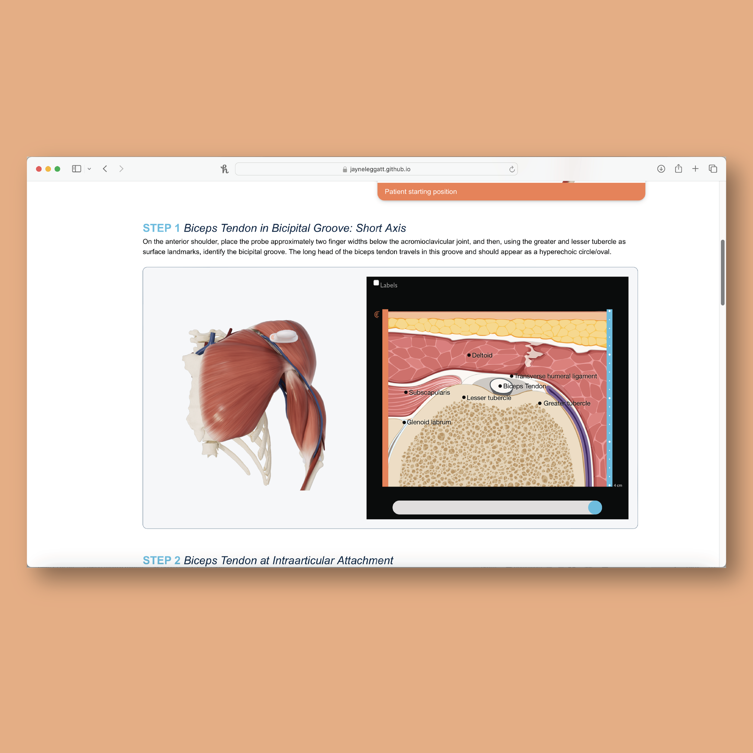
Sample Pages
Functional prototype coming in August!!


Research Poster
American Association for Anatomy - Anatomy Connected 2024
